 By
By
- Leslie P Gartner PhD Professor of Anatomy (Retired)Department of Biomedical Sciences
- James L Hiatt PhD Professor Emeritus, Department of Biomedical Sciences, University of Maryland Dental School, Baltimore, MD
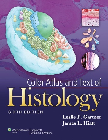
This best-selling atlas provides medical, dental, allied health, and biology students with an outstanding collection of histology images for all of the major tissue classes and body systems. This is a concise lab atlas with relevant text and consistent format presentation of photomicrograph plates. With a handy spiral binding that allows ease of use, it features a full-color art program comprising over 500 high-quality photomicrographs, scanning electron micrographs, and drawings. Didactic text in each chapter includes an Introduction, Clinical Correlations, Overview, and Chapter Summary.
Key Features
- NEW! Images added to the Clinical Considerations boxes.
- NEW! Larger trim size with larger photomicrographs.
- Clinical Considerations boxes in each chapter covering over 100 key conditions/topics.
- Concise text introducing and summarizing atlas plates with detailed legends and orientation thumbnail illustrations.
- Histophysiology text incorporated into the chapter Introductory material.
- Nearly 600 images, including photomicrograph atlas plates, electron micrographs, and schematic illustrations.
Download
Note: Only Gold member can download this ebook. Learn more here!








Dear admin
It would be great if you would open a forum for users to make some ebook requests (like internalmedicinebook.com).
Thanks for the upload.