 By
By
- Victor P. Eroschenko PhD, Professor of Anatomy, Department of Biological Sciences, University of Idaho WWAMI Medical Program, Moscow, ID
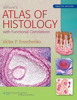
diFiore’s Atlas of Histology with Functional Correlations explains basic histology concepts through realistic, full-color composite and idealized illustrations of histologic structures. Added to the illustrations are actual photomicrographs of similar structures, a popular trademark of the atlas. All structures are directly correlated with the most important and essential functional correlations, allowing students to efficiently learn histologic structures and their major functions at the same time.
diFiore’s Atlas of Histology is the perfect resource for medical and graduate histology students.
New to This Edition
- Expanded Introduction on basic histology techniques and staining as well as a more comprehensive list of stains that students may encounter in their histology course
- New chapter on cell biology accompanied by both drawings and representative photomicrographs of the main stages in the cell cycle during mitosis
- Contents reorganized into four parts, progressing logically from Methods and Microscopy through Tissues and Systems
- Improved art program with digitally enhanced images to provide increased detail
- More than 40 new photomicrograph images, including light and transmission electron micrographs
- Student Resources: Online E-book, Interactive Question Bank for chapter review, and Interactive Atlas featuring all images from the book + more than 450 additional micrographs
Key Features
- NEW! Expanded Introduction including a new section that briefly describes the histology techniques, a new section on the cell cycle, and a more comprehensive list of different stains that students may encounter in examining the histology images during the course
- NEW! In-text references to the bonus online images
- NEW! Replacement/updating color of select micrograph-style illustrations
- NEW! Revised design to make the most use of space and showcase images
- NEW! Revised Table of Contents now includes 4 parts and updated chapter titles to more accurately reflect their contents
- NEW! Student ancillaries: Online e-book, Interactive question bank
- NEW! Updated chapter opening illustrations
- NEW! Updated functional information that pertains to different cells, tissues, and organs of the body that are illustrated in the atlas
- NEW! USMLE-style review questions and answers for each chapter (approximately 15 per chapter; approximately 315 for the book)
- Bold key terms for emphasis to help with learning and review
- Chapter opening overview illustrations (12) provide an overview of the tissue or organ being discussed
- Chapter summary outlines (bullets) for review
- Digitized full-color illustrations of histology slides (idealized for efficient learning)
- Faculty ancillaries: Image Bank (including all images in the book + additional micrographs)
- Full labels on images rather than abbreviations for faster learning
- Functional Correlations boxes (112) help students study structure and function together
- Over 70 micrographs adjacent to illustrations to provide a realistic perspective
- Student ancillaries: Interactive Atlas including all of the images in the book + more than 450 additional micrographs for reference and self-study
Download
Note: Only Gold member can download this ebook. Learn more here!
Note: This ebook is intended for promotional use only. We strongly recommend you to delete it from your computer within 24 hours. If you love this book, you should buy it’s newest Edition from Amazon.






Dear Admin
part 1 is damaged
Updated!
link not working