 By
By
- Stacey E Mills MD Professor of Pathology and Associate Director of Surgical Pathology, University of Virginia Health Sciences Center, Charlottesville, Virginia
- Darryl Carter
- Joel K Greenson MD Professor of Pathology, The University of Michigan Health System, Ann Arbor, MI
- Victor E Reuter MD Attending Pathologist, Memorial Hospital Member, Memorial Hospital, Memorial Sloan-Kettering Cancer Center; Professor of Pathology, Weill Medical College of Cornell University, New York, NY; Professor of Pathology, Weill Medical College of Co
- Mark H Stoler MD Professor of Pathology and Gynecology, Associate Director of Surgical Pathology and Cytopathology, University of Virginia Health System, Charlottesville, VA
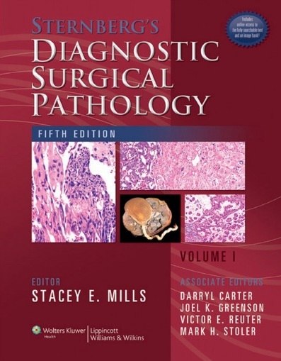
Completely updated, the Fifth Edition of this standard-setting two-volume reference presents the most advanced diagnostic techniques and the latest information on all currently known disease entities. More than 90 preeminent surgical pathologists offer expert advice on the diagnostic evaluation of every type of specimen from every anatomic site. The Fifth Edition contains over 4,400 full-color photographs.
This edition provides detailed coverage of the latest developments in the field, including new molecular and immunohistochemical markers for diagnosis and prognosis of neoplasia, improved classification systems for diagnosis and prognosis, the role of pathology in new diagnostic and therapeutic techniques, and the recognition of new entities or variants of entities. Where appropriate updated World Health Organization (WHO) terminology has been employed for tumor diagnosis. A few of the many specific changes include:
- An update on the serrated adenoma and serrated carcinoma pathway
- Virtually new chapters on salivary gland, larynx, blood vessels, heart, non-melanocytic skin tumors, and placenta
- Complete updates of the lymph node and bone marrow chapters incorporating the new WHO terminology
- An expanded discussion of the molecular biology of thyroid neoplasia
All full-color illustrations have been color-balanced to dramatically improve image quality.
A companion Website will include the fully searchable text and an image bank.
Key Features
- NEW: Detailed coverage of new developments in the field since publication of Fourth Edition, including:
- new molecular and immunohistochemical markers for diagnosis and prognosis of neoplasia
- improved classification systems for diagnosis and prognosis
- the role of pathology in new diagnostic and therapeutic techniques
- the recognition of new entities or variants of entities
- NEW: A few of the many content changes include:
- an update on the serrated adenoma and serrated carcinoma pathway
- virtually new chapters on salivary gland, larynx, blood vessels, heart, non-melanocytic skin tumors, and placenta
- complete updates of the lymph node and bone marrow chapters incorporating the new WHO terminology
- an expanded discussion of the molecular biology of thyroid neoplasia
- NEW: Updated World Health Organization (WHO) terminology for tumor diagnosis is used where appropriate
- NEW: All full-color illustrations have been color-balanced by the editor to dramatically improve image quality
- Provides latest diagnostic techniques
- Covers most diseases seen by surgical pathologists grouped by anatomical region
- Over 4,400 full-color illustrations spread through both volumes
- Over 90 expert contributors
Download
Note: Only Gold member can download this ebook. Learn more here!


