
 By
By
- Harald Kluge, MD, Former Professor and Head of the Central CSF, Laboratory, Faculty of Medicine and Institute of Clinical Chemistry, University of Jena, Jena, Germany
- Valentin Wieczorek †, Professor emeritus, Faculty of Medicine, Hans Berger Hospital of Neurology, University of Jena, Jena, Germany
- Ernst Linke, MD, Former Head of Central Laboratory, Currently Scientific supervisor of Central CSF, Laboratory, Asklepios Hospital, Stadtroda, Germany
- Klaus Zimmermann, MD, Head of Section for CSF Diagnostic Studies, Medical Laboratory Dresden/Elsterwerda, Laboratory Dr. Pontek/Dr. Bochmann, Dresden, Germany
- Stefan Isenmann, MD, Professor of Neurology, Supervising Physician, CSF Diagnostics, Department of Neurology, University Hospital Jena, Jena, Germany
- O. W. Witte, MD, Professor of Neurology, Head of Hans Berger Hospital of Neurology, University of Jena, Jena, Germany
A complete, single-volume reference for the cytological examination of cerebrospinal fluid!
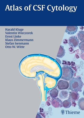
This full-color atlas presents all the essential information needed for reaching an accurate cytological diagnosis of cerebrospinal fluid and its abnormalities. Designed as a clinical and laboratory reference, Atlas of CSF Cytologyprovides an overview of all the standard diagnostic techniques and offers insight into advanced methods such as flow cytometry and immunocytological phenotyping. Brief descriptions of the indications, advantages, and limitations are provided for each method. An extensive collection of more than 300 high-quality cytological pictures demonstrating normal cell structures, as well as pathological cells in acute and remission phases enables the reader to understand disease processes.
Highlights:
- Guidelines for the proper handling of specimens, cell preparation, and staining techniques
- Review of the common sources of error in diagnosis
- Thorough coverage of the techniques for detecting and classifying inflammatory, infectious, neoplastic, and hemorrhagic conditions of the central nervous system
- Descriptions of the principle features of cells and the classification of tumor cell types according to current W.H.O. standards
- Full-color images depicting pathological alterations of CSF cells — an indispensable visual aid to comprehension
Atlas of CSF Cytology is ideal for specialists in neurology, neurosurgery, pathology/neuropathology, cytopathology, microbiology, and laboratory medicine, as well as for those internists, pediatricians, and psychiatrists who frequently request cytological examination of the CSF. Though it is written to meet the needs of specialists, the Atlas will also be found accessible and enlightening by interested medical students, interns and residents.
Download
Note: Only Gold member can download this ebook. Learn more here!
Related Books
 Gynecologic Cytology
 Atlas of Anatomy, 3rd Edition Latin Nomenclature
 Color Atlas of Pathophysiology, 2nd Edition
 Pocket Atlas of Human Anatomy Founded by Heinz Feneis, 5th Edition
 Color Atlas of Genetics, 4th Edition
 Color Atlas of Pathophysiology, Third Edition
 Thurlbeck’s Pathology of the Lung, 3rd Edition
 Color Atlas of Human Anatomy, Vol 2: Internal Organs, 5th Edition
 Atlas of Anatomy, 2nd Edition
 Pharmacology – An Illustrated Review
Dear Admin
Thanks for this book!!
Can you upload please if you have in your collection
Color Atlas of Differential Diagnosis in Exfoliative and Aspiration Cytopathology by Lippincott Williams & Wilkins; Second edition (May 2011)
Disappear for download.Can you try re-upload or new edition.
New link added!